3D Systems offers both 3D printers and software for creating FDA-cleared, diagnostic-quality anatomic models. You can select from a broad range of materials capable of biocompatibility and sterility to 3D print on technologies that include MultiJet Printing, Stereolithography and Selective Laser Sintering.
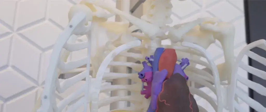
Since each patient's anatomy is different, patient-specific anatomic models provide an opportunity to visualize and plan surgeries prior to entering the operating room. In addition, you can use anatomic models to educate patients and their families on an upcoming procedure.

Models printed in flexible, rigid, opaque and clear materials

Translucent and opaque models printed in our high-strength materials*

Strong, durable models in medical-grade nylon materials*
*Materials capable of biocompatibility and sterility
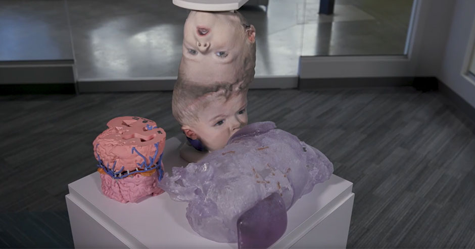
The remarkable achievement of successfully separating conjoined twins, Pedro and Augusto, presented a daunting challenge. Arriving at Dayton Children’s Hospital in July 2021, these boys, nearing their third birthday, arrived from Guatemala. They were conjoined at the top of their heads, positioned at a 90-degree angle, a condition classified as craniopagus O’Connell class III.
The documentary provides a comprehensive account of the two-year journey undertaken by surgeons and medical staff. Their meticulous preparation and execution of a series of intricate separation surgeries underscore the complexity of this rare medical circumstance.

You can count on 3D Systems for a broad range of anatomic model solutions—from design to delivery. Whether you are looking for a way to create a digital 3D model from DICOM, you already have a design file and need a printing service, or you need an end-to-end solution, we have you covered. Solutions include:

MultiJet printing quality, speed and ease of use made accessible

Industrial-scale, process controlled additive manufacturing solution for selective laser sintering